Leg Bone Diagram : Premium Vector Anatomy Of Human Knee Sketch Of Leg Bones And Joint Medicine Side And Front View Of Knee Bones Hand Drawn Femur Patella Tibia And Fibula Tibial Plateau And : The hip itself is a ball and socket joint, much like the shoulder.the structures necessary to create this joint are the socket, the joint capsule, muscle, ligaments, and the neck.
Leg Bone Diagram : Premium Vector Anatomy Of Human Knee Sketch Of Leg Bones And Joint Medicine Side And Front View Of Knee Bones Hand Drawn Femur Patella Tibia And Fibula Tibial Plateau And : The hip itself is a ball and socket joint, much like the shoulder.the structures necessary to create this joint are the socket, the joint capsule, muscle, ligaments, and the neck.. Inflammation of navicular bone and/or bursa. Related posts of leg bones anatomy diagram cross section of foot nerves. Its decrease finish helps create the knee joint. Sometimes the fractured bone fails to heal in the proper position. The lower extremity, commonly referred to as the leg, contains four bones (the femur, the patella, the tibia, and the fibula) and bends at the hip, the knee, and the ankle.
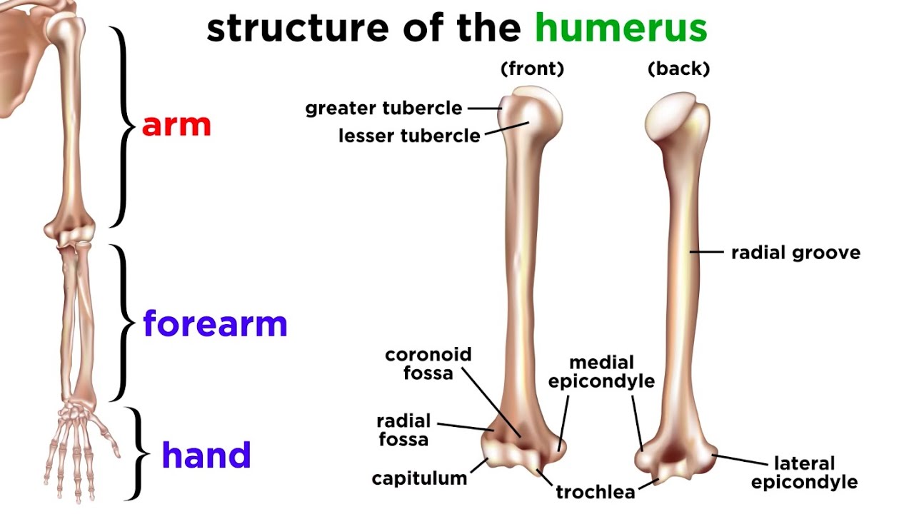
Full muscles of the leg medical edition 3d model | cgtrader. Click now to learn more about the bones, muscles, and soft tissues tibia: The lower leg extends from the knee to the ankle. File human arm bones diagram svg wikipedia. The thigh bone, or femur, is the large upper leg bone that connects the lower leg bones (knee joint) to the pelvic bone (hip joint).
Practical Art Anatomy E G Lutz from drawingbooks.org Anatomy of the foot (26/28 bones) 11 terms. Its lower end helps create the knee joint. Tibia and fibula the tibia and fibula are two long bones that run parallel to each other, forming the scaffold of the leg and providing attachment points for many muscles. These landmarks are the anterior superior iliac spine. Click now to learn more about the bones, muscles, and soft tissues tibia: Distal end of right humerus. The lower leg extends from the knee to the ankle. The lumbar plexus forms in the lower back from the merger of spinal nerves l1 through l4 while the.
The tibia and the fibula, at the top of the ankle joint.
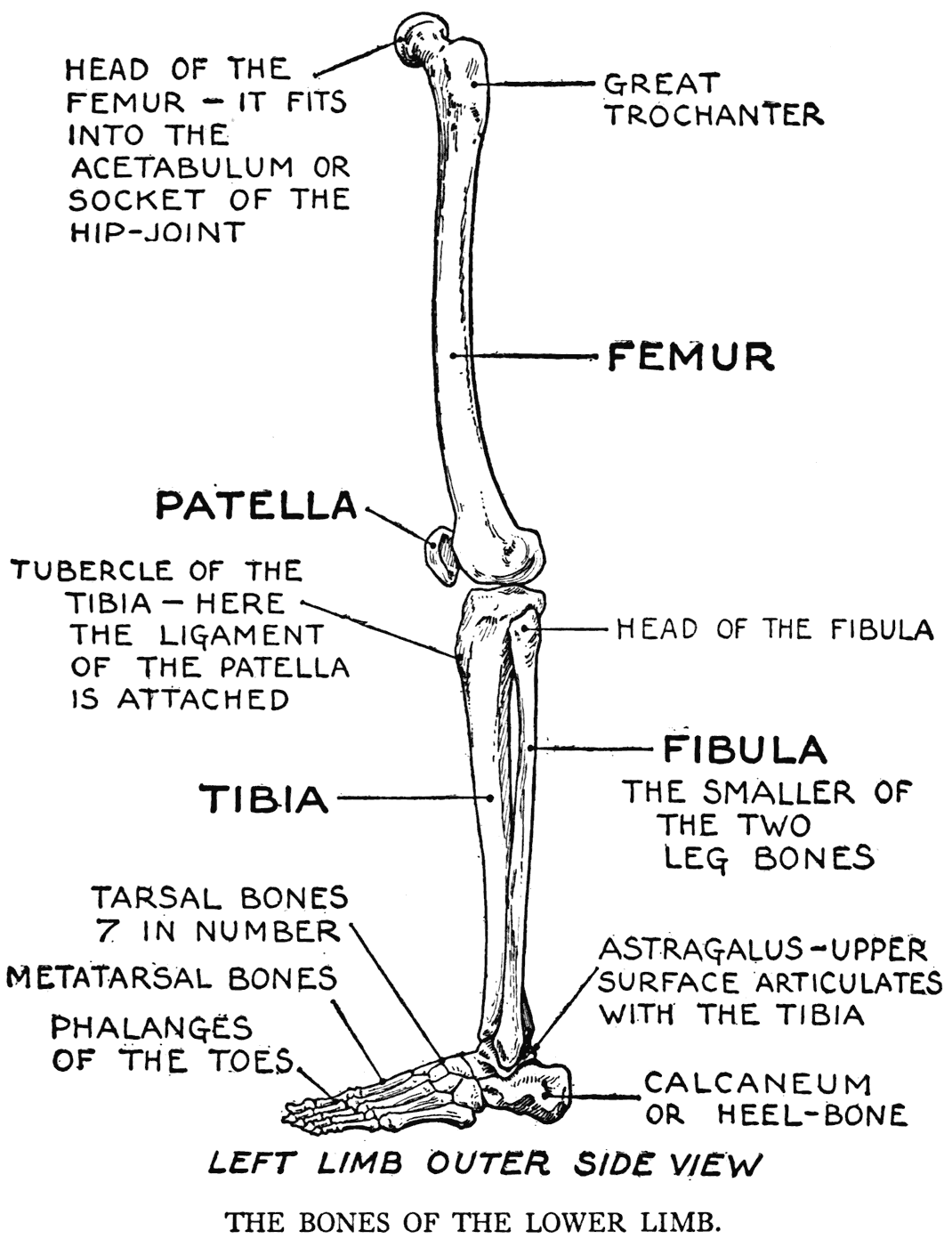
Another bone that is part of the lower leg and the knee joint is called the fibula.this is a bone located on the lateral, or outer part, of the lower leg and is more commonly known as the calf bone. The largest and most medial leg bone, forming both the knee and ankle joints. The femur, or thighbone, is the longest and largest bone in the human body. Its decrease finish helps create the knee joint. As these nerves descend toward the thighs, they form two networks of crossed nerves known as the lumbar plexus and sacral plexus. The lower leg is comprised of two bones the tibia and the smaller fibula. Cross section of foot nerves 13 photos of the cross section of foot nerves cross section of nerve fiber, foot anatomy nerves, foot nerve pain, human foot nerves, nerve cross section histology, peripheral nerve cross section, spinal nerve cross section, foot, cross section of nerve fiber, foot anatomy nerves, foot. The femur is the single bone of the thigh. Anatomy of the foot (26/28 bones) 11 terms. File human arm bones diagram svg wikipedia. This diagram depicts diagram leg bones anatomy.human anatomy diagrams show internal organs, cells, systems, conditions, symptoms and sickness information and/or tips for healthy living. Poster | zazzle.com (ralph chavez) The major bones of the leg are the femur (thigh bone), tibia (shin bone), and adjacent fibula, and these are all long bones.the patella (kneecap) is the sesamoid bone in front of the knee.most of the leg skeleton has bony prominences and margins that can be palpated and some serve as anatomical landmarks that define the extent of the leg.
As these nerves descend toward the thighs, they form two networks of crossed nerves known as the lumbar plexus and sacral plexus. This area is commonly referred to as the calf. The femur, or thighbone, is the longest and largest bone in the human body. This muscle runs along the outside of the back of your thigh and attaches to the top of the fibula (the smaller of the two bones of your lower leg). Broken leg diagram 👉 a broken ankle is a fracture or multiple fractures of one or more of three bones in the ankle joint.
The Skeletal System Youtube from i.ytimg.com Inflammation of navicular bone and/or bursa. Great for halloween, or anytime of the year. Diagramme schnell und einfach erstellen. The femur, or thighbone, is the longest and largest bone in the human body. The bones of the leg are the femur, tibia, fibula and patella.the foot bones shown in this diagram are the talus, navicular, cuneiform, cuboid, metatarsals and calcaneus. The leg is specifically the region between the knee joint and the ankle joint. The lower leg is comprised of two bones the tibia and the smaller fibula. Related posts of leg bones anatomy diagram cross section of foot nerves.
The hip itself is a ball and socket joint, much like the shoulder.the structures necessary to create this joint are the socket, the joint capsule, muscle, ligaments, and the neck.
The largest and most medial leg bone, forming both the knee and ankle joints. Diagramme schnell und einfach erstellen. The tibia, commonly known as the 'shin bone', is the largest and most medial of the two.you can palpate its anterior border when you run your finger down the anterior aspect of your leg. Inflammation of navicular bone and/or bursa. The femur, or thighbone, is the longest and largest bone in the human body. Cross section of foot nerves 13 photos of the cross section of foot nerves cross section of nerve fiber, foot anatomy nerves, foot nerve pain, human foot nerves, nerve cross section histology, peripheral nerve cross section, spinal nerve cross section, foot, cross section of nerve fiber, foot anatomy nerves, foot. Performance horses tend to suffer from this degenerative disease. They support the legs to bear the body weight and also help in proper locomotion. With different grades of sprains depending on severity. Click now to learn more about the bones, muscles, and soft tissues tibia: The lumbar plexus forms in the lower back from the merger of spinal nerves l1 through l4 while the. The lateral and smaller bone of the lower leg. The lower extremity, commonly referred to as the leg, contains four bones (the femur, the patella, the tibia, and the fibula) and bends at the hip, the knee, and the ankle.
The lumbar plexus forms in the lower back from the merger of spinal nerves l1 through l4 while the. The lower limb contains 30 bones. Master leg and knee anatomy using our topic page. The pubis, ischium, and ilium together constitute the pelvis while the thigh bone is the femur. Human leg bone diagram :
Anatomy Chart Of Human Bones For Stock Vector Colourbox from d2gg9evh47fn9z.cloudfront.net Inflammation of navicular bone and/or bursa. The lateral and smaller bone of the lower leg. Diagramme schnell und einfach erstellen. This type of fracture and its classification system is named for robert b. Poster | zazzle.com (ralph chavez) The human leg consists of 8 bones, 4 per leg. The femur, or thighbone, is the longest and largest bone in the human body. Cross section of foot nerves 13 photos of the cross section of foot nerves cross section of nerve fiber, foot anatomy nerves, foot nerve pain, human foot nerves, nerve cross section histology, peripheral nerve cross section, spinal nerve cross section, foot, cross section of nerve fiber, foot anatomy nerves, foot.
It is likely that abnormal biomechanical stresses are the basis for the disease.
The bones of the leg are the femur, tibia, fibula and patella.the foot bones shown in this diagram are the talus, navicular, cuneiform, cuboid, metatarsals and calcaneus. Bones of foot, labeled diagram. The bones together make up the hip. This area is commonly referred to as the calf. Poster | zazzle.com (ralph chavez) Inside of arm muscle and bone 12 photos of the inside of arm muscle and bone , bone Related posts of leg bones anatomy diagram cross section of foot nerves. Great for halloween, or anytime of the year. The lower leg is comprised of two bones the tibia and the smaller fibula. Its lower end helps create the knee joint. Degenerative disease, similar to arthritis. Its decrease finish helps create the knee joint. The nerves of the leg and foot arise from spinal nerves connected to the spinal cord in the lower back and pelvis.


0 Komentar